Cystotomy and Scrotal Urethrostomy
Cystic calculi, also called bladder stones or uroliths, are commonly seen in both dogs and cats. There are four predominant types of bladder stones: struvite, calcium oxalate, purine, and urate. Between 5-15% of bladder stones are mixed composition. Treatment options and risk factors depend on the type of stone present, therefore each bladder stone is outlined below:
- Struvite
Struvite stones are composed of magnesium ammonium phosphate hexahydrate (figure 1A & B).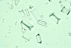
Figure 1A. Struvite crystals in urine sediment. Photo courtesy of Meyer D, Raskin RE, Atlas of Canine and Feline Cytology, Saunders, 2001.
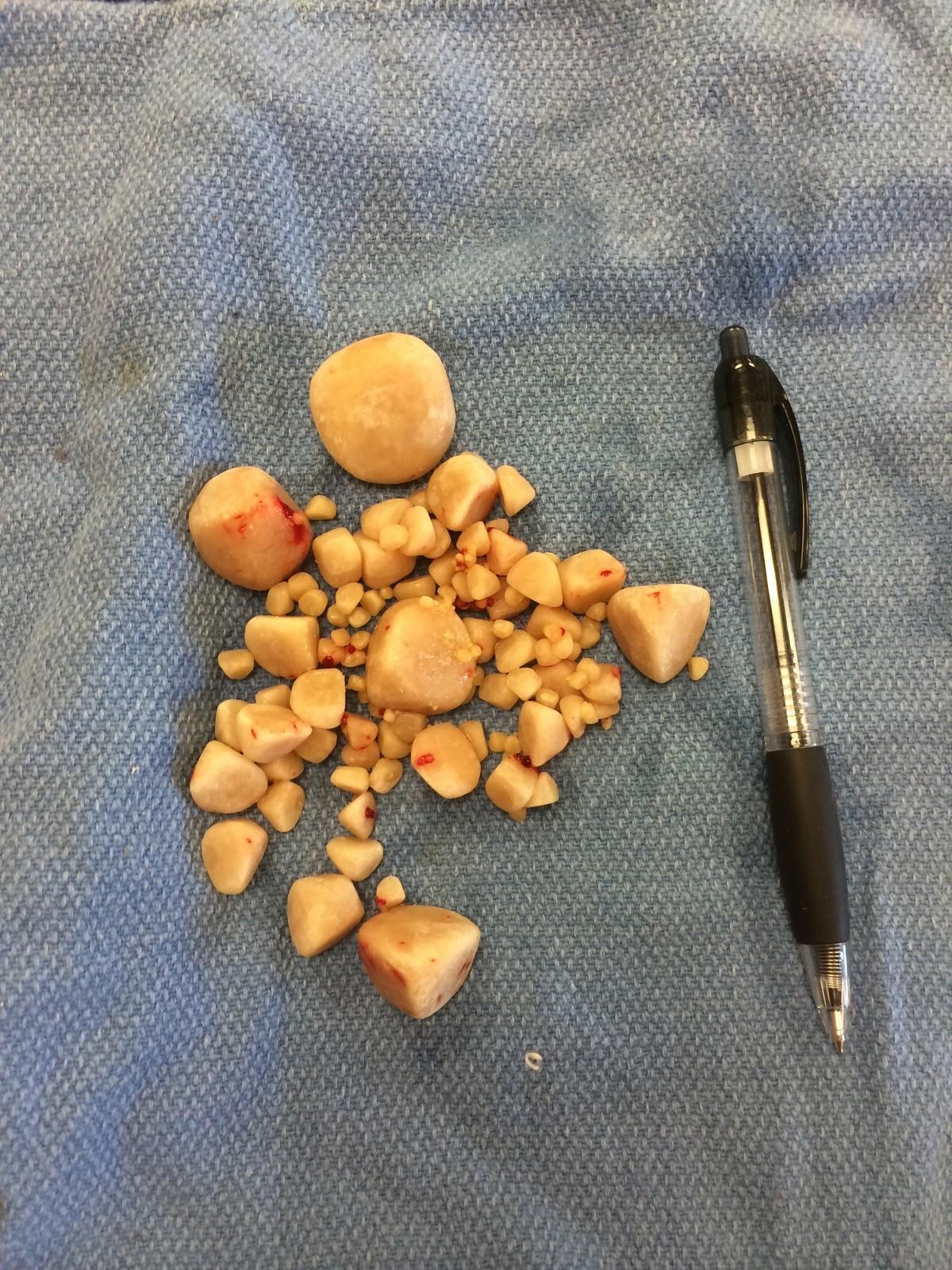
Figure 1B. Struvite stones. Photo courtesy of Dr. Joanne Franks, Dallas Veterinary Surgical Center.
Struvite stones are the most common bladder stone in dogs, and are often associated with urinary tract bacterial infections. Cats more commonly have sterile struvites, meaning the stones are not associated with a urinary tract infection. Since female dogs have an increased risk of urinary tract infections, struvite stones occur most commonly in female dogs. Struvite stones can be dissolved by a struvite dissolution diet +/- administering an appropriate antibiotic based on urine culture and sensitivity. This dissolution process can take weeks to months.
- Calcium oxalate
Calcium oxalate stones can be associated with aciduria (acidic urine) and hypercalcemia (elevated blood calcium levels) (figure 2). Certain breeds of cats and dogs are predisposed, such as long-haired cats and small breeds of dogs (miniature schnauzer, Lhasa Apso, Shih Tzu, and Yorkshire terrier breeds). Unfortunately, calcium oxalate stones cannot be dissolved via medial management.
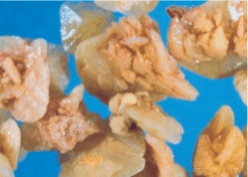
Figure 2. Calcium oxalate stones. Photo courtesy of Dr. Gregory F Grauer, DVM, MS, DACVIM, Calcium Oxalate Urolithiasis, Clinician’s Brief 2014.
- Purine
Purine stones or urates are made up of 3 groups of compounds: oxypurines, aminopurines, and methylpurines (figure 3).
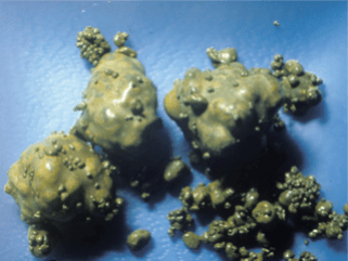
Figure 3. Urate Stones. Photo courtesy of V. Ling Urinary Stone Analysis Laboratory, UC Davis, http://www.vetmed.ucdavis.edu/usal/veterinarians/stone_management_sheets.cfm.
These stones are more common in dogs than cats. They are often seen in animals less than 7 years of age. Urates typically form secondary to liver disease such as portosystemic vascular shunts. Certain breeds of dogs such as Dalmatians or English bulldogs are predisposed to metabolic disorders which cause an elevated level of urate in the urine. It is possible to dissolve urate stones with a purine restricted diet and medications. However if an underlying condition exists, such as liver disease, this must be treated first.
- Cystine
Cystine stones are commonly seen in dogs that have a genetic defect. They are usually seen in Newfoundlands, Dachshunds, Basset hounds, English Bulldogs, Chihuahuas, Yorkshire Terriers, Irish Terriers, and mixed-breed dogs. It is seen most commonly in male dogs. Canine cystine stones can be dissolved medically, but this has been unsuccessful in cats.
Symptoms
The most common symptoms associated with bladder stones include stranguria (straining to urinate), pollakiuria (urinating small and frequent amounts), hematuria (blood in the urine), or inappropriate urination (urinating in the house). These clinical signs can be the same for urinary tract infections, therefore diagnostics to rule out both of these conditions is recommended. Occasionally, a bladder stone can migrate from the bladder and obstruct the urethra. Clinical signs of a bladder obstruction include straining to urinate with minimal urine production, vocalizing, vomiting, decreased appetite, and lethargy. Animals unable to urinate can develop severe electrolyte abnormalities, changes in blood pressure, heart arrhythmias, kidney failure, and even death. Urinary obstruction is considered an emergency and immediate treatment by a veterinarian is essential.
Diagnosis
Abdominal radiographs (x-rays) are the most definitive diagnostic tool for detection of most uroliths (figure 4 + 5A).
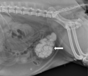
Figure 4. Radiograph (x-ray) of a canine bladder with multiple uroliths.
Photo courtesy of Dr. Joanne Franks, Dallas Veterinary Surgical Center.
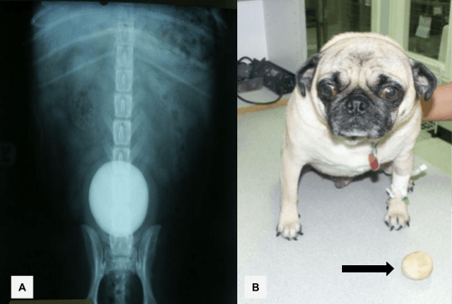
Figure 5. A) Radiograph (x-ray) of a canine bladder with a single large bladder stone. B) The bladder stone next to the dog after surgical removal.
Photos courtesy of Dr. Joanne Franks, Dallas Veterinary Surgical Center.
However, not all stones can be seen on radiographs. Therefore ultrasonography or double contrast cystography may be necessary. Contrast cystography is a medical dye study used to highlight radiolucent stones on x-rays.
Additional diagnostics that are recommended include urinalysis and urine culture. A urinalysis can provide information regarding urine specific gravity and urine pH which can suggest what type of urolith is present. A urinalysis also informs the veterinarian if crystalluria is present. While crystalluria can suggest crystalline oversaturation this does not confirm or deny the presence of bladder stones. As previously discussed, some bladder stones such as struvite stones can be associated with bladder infection. Therefore a urine culture and sensitivity will give us information regarding the presence of a urinary tract infection as well as the appropriate antibiotics to treat the infection.
Finally, bloodwork is often indicated. This is to rule out the presence of other disease processes such as hypercalcemia (elevated blood calcium levels) or liver disease which can predispose animals to certain types of bladder stones.
Treatment – medical vs surgical
If the type of stone is known and it is amenable to dissolution, medical management can be attempted. However often times we do not know the type of stone present or if it is a type that can be dissolved. Therefore, surgery will be recommended.
Surgery
The most common surgical procedure for cystic calculi is a cystotomy. For this procedure an incision is made into the abdominal cavity. The bladder is located and isolated (figure 6A).
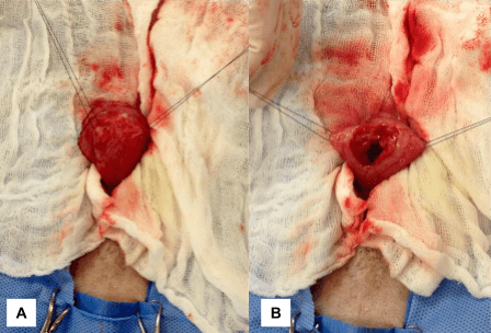
Figure 6. A) The bladder is isolated outside of the abdominal cavity and held in place with sutures. B) An incision is then made into the bladder. Photos courtesy of Dr. Joanne Franks, Dallas Veterinary Surgical Center.
A small incision is then made into the bladder (figure 6B). The stones are identified and removed (figure 7A). Often a catheter is placed from the urethra to the bladder and then vice versa to flush any remaining stones that could have migrated into the urethra. Once all the stones are removed, the bladder wall is sutured back together followed by closure of the abdominal wall (figure 7B).
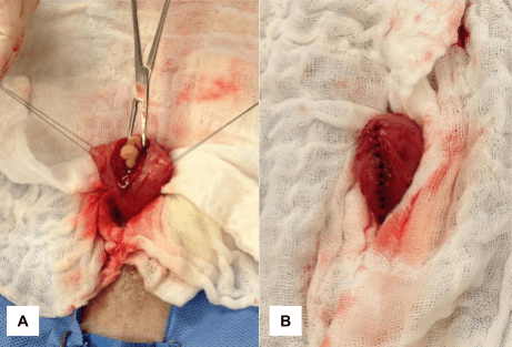
Figure 7. A) The bladder stone is identified and removed. B) Once all the stones are removed the bladder wall is sutured closed. Photos courtesy of Dr. Joanne Franks, Dallas Veterinary Surgical Center.
Occasionally a sample of the bladder mucosa may be submitted for culture and sensitivity. Radiographs may be performed after the procedure to confirm removal of all bladder stones.
There are other surgical procedures that may be considered with cystic calculi such as prescrotal urethrostomy, scrotal urethrostomy, and perineal urethrostomy. Scrotal urethrostomy will be explained in further detail below.
Postoperative care
Typically, animals stay one night in the hospital for monitoring during their recovery. It is imperative that the animal wears an e-collar at all times to prevent licking, self-trauma, and damage to the surgical repair until healed. Two to three weeks of exercise restriction is typically recommended to allow the bladder to heal. Pain medication, anti-inflammatories, and antibiotics are prescribed at the surgeon’s discretion. Depending on the suture material used, follow-up may be required for suture removal. Additional medical management may be warranted based on stone analysis and/or culture and sensitivity. Animals may continue to strain to urinate, have frequent urination, or hematuria (blood in the urine) for 3-4 days after surgery.
Potential complications
Complications from this procedure are rare. Possible complications include incision dehiscence, leakage, infection, stricture/granulation tissue formation, or missing a stone in the bladder during surgery.
Prognosis
The prognosis for a cystotomy is excellent. However, it is important to note that depending on the type of calculi present, stone recurrence can occur. Recommendations in terms of medical management and stone prevention will be made after stone analysis.
Scrotal Urethrostomy
As previously mentioned, a bladder stone can occasionally migrate and obstruct the urethra. This occurs more commonly in male dogs and cats because their urethra is anatomically smaller than in females. Given that this is a surgical emergency, an additional surgical procedure may be recommended to prevent the occurrence of a urethral obstruction in male dogs with multiple episodes of bladder stones. The most common surgical procedure in male dogs is known as a scrotal urethrostomy. Please see the additional DVSC page on Perineal Urethrostomy to read about the most common surgical procedure in male cats.
A scrotal urethrostomy is a surgically created new opening in the urethra to bypass the narrow area where obstruction usually occurs. An incision is made through the skin and urethra at the level of the scrotum (figure 8A & 8B). The lining of the urethra is then sutured to the skin (figure 8C).
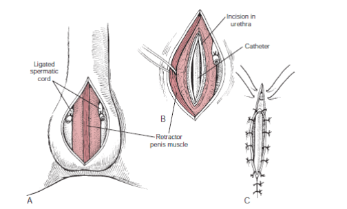
Figure 8. A) A skin incision is made into the scrotum. B) A second incision is made into the urethra. C) The urethral lining is then sutured to the skin to create a larger urethral opening. Photo courtesy of Fossum: Small Animal Surgery 3rd edition<br />Copyright © 2007 by Mosby, Inc., an affiliate of Elsevier Inc.
This procedure results in a wider opening of the urethra for small stones to pass. It is important to note that if the animal is intact they will have to be neutered to undergo this procedure. Possible post-operative concerns include hemorrhage (bleeding at the surgical site for a few days to weeks), recurrent urinary tract infections (15% of cases), intermittent urine irritation of the skin, having to keep hair trimmed around the new opening, and rarely recurrent obstruction from urinary stones. However, most dogs do very well this procedure. Postoperative care and recovery are similar to those above for a cystotomy.
Megan Cray, VMD
Comments Closed