Home » Medical Library » Urethral Prolapse
Urethral Prolapse
Urethral Prolapse
Urethral prolapse is defined as the extrusion of the urethral mucosa through the external urethral orifice of the penis. This condition occurs most commonly in young male dogs that have not been neutered. Brachycephalic breeds (e.g., bulldogs, Boston terriers, pugs) and Yorkshire terriers may be predisposed to urethral prolapse.
Although we don’t know why this condition occurs, possible causes include increased intra-abdominal pressure secondary to upper airway obstructive syndrome (common condition in brachycephalic breeds), dysuria (painful or difficult urination), developmental abnormalities, or sexual excitement. Urethral prolapse has been reported to be secondary to cystitis (inflammation within the bladder). Therefore, additional diagnostics may be warranted.
Symptoms and Diagnosis
The most common clinical signs that will be seen at home are excessive licking of the penis, blood in the urine, or bleeding from the prepuce or tip of the penis.
The diagnosis of urethral prolapse is made on physical examination with a veterinarian. Upon extrusion of the penis from its prepuce, a red to purple mass is observed at the distal end of the penis. This mass is the inflamed protruded urethral mucosa (figure 1).
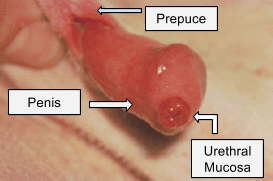
Figure 1. A prolapsed urethra. Photo courtesy of Fossum: Small Animal Surgery 3rd edition<br />Copyright © 2007 by Mosby, Inc., an affiliate of Elsevier Inc.
As previously discussed, urethral prolapse can be a result of cystitis (inflammation of the bladder). Cystitis can be secondary to a variety of causes including urinary tract infections or uroliths (urinary tract stones). Therefore, abdominal radiographs may be recommended to rule out the presence of urinary stones. It may also be recommended that a sterile sample of urine be obtained via cystocentesis. This procedure requires inserting a needle into the bladder through the abdomen to obtain urine. This sample can then be submitted for urine culture and sensitivity to rule out a urinary tract infection. It will be determined during your initial surgical consult as to whether or not these diagnostics are warranted.
Treatment – medical vs surgical
With no treatment the urethral prolapse will not resolve on it’s own. Manual reduction of the prolapsed segment can be attempted if the tissue protrusion is minimal or if the animal is asymptomatic. This procedure requires general anesthesia. Once the urethral tissue is manually reduced (pushed back inside), a purse-string suture pattern is placed in the tunic of the penis at the external urethral orifice. This suture is maintained for approximately 5 days before being removed. Recurrence with this procedure is common. Therefore, surgery is often recommended. If a dog has failed conservative management or if they present with excessive bleeding, discomfort, or a severely damaged urethral mucosa, surgery is strongly recommended.
Surgery
There are two types of surgical procedures that can be performed to treat urethral prolapse. The first is a resection and anastomosis of the urethral mucosa. With this procedure, the urethral mucosa is gently retracted, and the affected urethra tissue is transected (figure 2A). The remaining viable tissue is then sutured to the penis (figure 2B and figure 3).
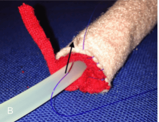
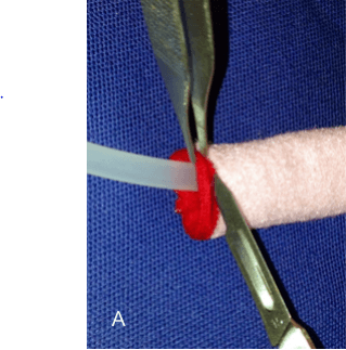
Figure 2. A. Surgical model demonstrating resection of a urethral prolapse. B.Surgical model demonstrating suturing the mucosa to the penis. Photo courtesy of ST Birchard. Veterinary Key Points. URL: http://drstephenbirchard.blogspot.com/2014/09/urethral-prolapse-in-dogs-why-it.html
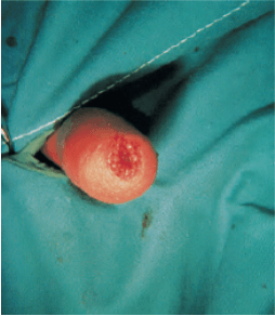
Figure 3. Final appearance of the penis after resection and anastomosis. Photo courtesy of Fossum: Small Animal Surgery 3rd edition<br />Copyright © 2007 by Mosby, Inc., an affiliate of Elsevier Inc
Alternatively, a urethropexy is an additional surgical procedure that is commonly used (figure 4). The penis is manually extended from the prepuce (4A). A grooved director is then introduced into the urethra which reduces the prolapse (4B). Suture is then passed full thickness through the penis proximally to the intraluminal surface out of the urethral orifice (4C). The needle is then passed in the opposite direction from the urethral lumen to the external surface of the penis (4D). A knot is then tied with the resulting full thickness sutures (4E). This is repeated until 2-4 equally spaced sutures are placed.
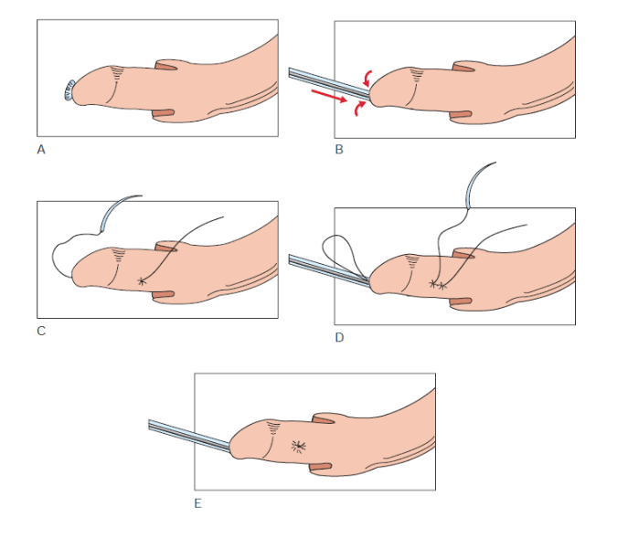
Figure 4. Urethropexy procedure. Photo courtesy of Fossum: Small Animal Surgery 3rd edition<br />Copyright © 2007 by Mosby, Inc., an affiliate of Elsevier Inc
With either surgical procedure, it is strongly recommended that castration be performed at the same time because sexual arousal is thought to be a contributing factor in urethral prolapse. Castration may help prevent recurrence.
Potential complications
Regardless of the procedure used, complications include continuous hemorrhage or bleeding, penile swelling, stranguria (straining to urinate), and prolapse recurrence. Prolapse recurrence with surgery has been reported to occur in 57% of cases. It is expected that there will be intermittent hemorrhage or bleeding postoperatively particularly during urination for a few days after surgery.
Postoperative Care
Typically, animals stay one night in the hospital for monitoring during their recovery. It is imperative that for 2 weeks after surgery the animal wears an e-collar at all times to prevent licking, self-trauma, and damage to the surgical repair. Two to three weeks of exercise restriction is typically recommended to allow the urethra to heal. Pain medication, anti-inflammatories, and antibiotics are prescribed at the surgeon’s discretion. Depending on the suture material used, follow-up may be required for suture removal.
Prognosis
The vast majority of owners are pleased with the ultimate surgical outcome and are satisfied with the procedure. However, it is important to note that urethral prolapse recurrence does occur in a moderate number of cases, and a second surgical procedure may be necessary. Postoperative care such as wearing an e-collar and keeping an animal quiet after surgery may help prevent recurrence.
Author: Megan Cray, VMD
Comments Closed