Home » Medical Library » The Facts About Backs (IVDD)
The Facts About Backs (IVDD)
Intervertebral disc disease (IVDD) is a very serious and common disease seen in thousands of dogs every year. IVDD most frequently affects chondrodystrophic dogs—those characterized by having short legs and long backs, like the Dachshund and Bassett hound for example. However, IV disc herniation can occur in any breed—even in cats.
Anatomy and disease process
The spine is made up of individual interconnected bones called vertebra. The vertebral column extends from the back of the skull to the tail, and is divided into sections: cervical (neck), thoracic (upper back, with ribs attached), lumbar (lower back, no ribs), sacrum, and caudal (tail). Each vertebra has a hole through which the spinal cord traverses the length of the spine. Between each vertebra are spinal nerves, which control body functions. Just below the spinal cord and between each vertebra is the intervertebral disk. The disk is made up of two parts, a soft pliable center and a tough flexible capsule. See Figure 1.

Figure 1. Normal canine spinal anatomy. From Hill’s Pet Nutrition, from the Atlas of Veterinary Clinical Anatomy, http://www.hillsvet.com/practice-management/vertebrae-intervertebral-disk-disease.html
In dogs with intervertebral disk disease (IVDD), the center of the disk becomes calcified and the capsule becomes brittle. The capsule of the disk can rupture without warning, allowing the calcified material in the center of the spinal cord to enter the spinal canal and put pressure on the spinal cord (Figure 2).

Figure 2. Illustration of intervertebral disk herniation. From Hill’s Pet Nutrition, from the Atlas of Veterinary Clinical Anatomy, http://www.hillsvet.com/practice-management/vertebrae-intervertebral-disk-disease.html
This disk rupture can happen acutely, causing signs of pain and paralysis within a matter of hours (a Type 1 rupture), or it can happen slowly over time, causing chronic low grade back pain and nerve damage (a Type 2 rupture). Some dogs have a combination of both types. Common terms for IVDD include “disk rupture” or “slipped disk.”
Symptoms
Without a doubt, IVDD is the most common neurosurgical condition presented to veterinarians. Herniated discs occur in the cervical (neck) and thoracolumbar spine (lower back). When a disk ruptures and puts pressure on the spinal cord, the signals from the brain to the legs are not transmitted normally.
If a dog ruptures a disk in the neck, all four legs are affected. If a dog ruptures a disk in the back, the front limbs will have normal function, but the back legs will be affected.
The severity of the symptoms a dog has is directly related to how much spinal cord damage has occurred. A small disk rupture, or one that happens slowly and gradually over several weeks, may only cause back or neck pain with minimal paralysis. If a disk ruptures fast and forcefully, the pressure on the spinal cord will cause pain and varying degrees of paralysis.
When a dog experiences acute Type 1 disc disease (extrusion of the center of the disk) a range of symptoms may be seen. With mild disease, back pain and ataxia (walking wobbly or unsteadily in the rear limbs) can be seen. More significant pressure on the spinal cord will cause loss of the placing reflex or inability to right the toes if they are curled under when standing (called conscious proprioception), Figure 3. In more severe cases, complete paralysis (loss of ability to move the legs) and even loss of pain sensation to the feet may occur.
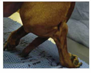
Figure 3. Photograph of a dachshund that has lost the ability to correct the position of the back foot, called loss of proprioception.
Progression of symptoms of IVDD occur in an orderly fashion:
Stage 1: Neck or back pain without neurological deficits
Stage 2: The ability to walk but with proprioception deficits (knuckling of paws, figure 3), and incoordination (ataxia or paresis)
Stage 3: The ability to move the legs, but inability to stand and walk under their own power
Stage 4: Paralysis, which is the complete inability to move the legs but maintaining the ability to feel a deep pinch of the toes
Stage 5: Paralysis with no feeling of a deep pinch to the toes
These signs always worsen in the order listed above, and they always return in the reverse order in the recovering patient. A dog that can walk has to have pain sensation to the feet, and a dog that cannot feel its toes cannot possibly be walking.
Reflexes
Patellar, sciatic, panniculus, and withdrawal (pulling back the foot when pinched) are all examples of reflexes, controlled by a lower motor neuron arc in the spinal cord. Reflexes are commonly misinterpreted during a neurological exam.
The presence or absence of spinal reflexes only helps the veterinarian to localize the lesion to a specific region of the spinal cord. Reflexes do not indicate the degree of cord damage or prognosis.
Dogs can have complete disruption of spinal cord function and still have reflexes, particularly withdrawal from a toe pinch. Therefore, withdrawal of the limb is NOT an accurate assessment of neurologic stage as defined above, or prognosis.
Assessment of pain sensation
Pain sensation (commonly called “deep pain”) is used to assess stage 4 and 5 (as listed above) because stage is closely correlated with prognosis. Pain perception is tested in animals by pinching the bone of a toe with hemostats. It is not necessary to pinch the toes of dogs with Stage 1, 2, or 3 IVDD.
Like reflexes, pain perception is commonly misinterpreted during a neurological exam. Pain perception is present or positive in a pet only if there is a conscious mental response to the toe being pinched, like turning the head toward the pinch, crying out, trying to bite the pincher, or trying to get away, Figure 4 (video). If the dog pulls its leg back, but it doesn’t act like it is being hurt, deep pain is NOT present. Simply pulling the leg back in response to a pinch is simply a withdrawal reflex, Figure 5 (video).
Figure 4 (above). Video demonstration of the presence of deep pain, positive sensory, Stage 4. Note that the patient demonstrates a mental response to being pinched, proving that the painful stimulus is in fact felt.
Figure 5 (above). Video demonstration of the absence of deep pain, negative sensory status, Stage 5. Note that a withdrawal reflex (the dog pulling back the leg) is still intact, which is common, but does NOT mean that deep pain is present.
Diagnosis
Diseases such as trauma, fibrocartilaginous emboli (FCE), meningitis, inflammatory disease, infection, and cancer can mimic the symptoms of disc herniation. Several tests can be performed to determine the underlying cause of pain or paralysis.
Diagnostic spinal imaging techniques used to diagnose disc herniation include radiography (xray), myelogram, CT scan, or MRI. Each form of imaging has its own unique benefits and pitfalls.
Plain radiographs (Figure 6) of the spine are useful in evaluating the bony spinal column for position of bones, continuity of the spinal column and spinal canal, bone density, and integrity of vertebral body end plates and disc spaces. Correct positioning and radiographic technique are critical for evaluating the spine.
Lesions that could possibly be diagnosed with plain radiographs of the spine include spinal fractures, luxations, vertebral anomalies (such as hemivertebrae or spina bifida), discospondylitis (disk infection), and some vertebral cancers.
However, because the spinal cord and discs cannot be seen on plain radiographs, xrays alone are inadequate to diagnose intervertebral disc herniation or to plan surgical intervention, and are of limited benefit in the patient with IVDD.

Figure 6. Example of a plain (noncontrast) radiograph of the thoracolumbar spine.
Computed Tomography (CT scan) (Figures 8 and 9) is an excellent imaging technique for IVDD. A CT scan involves the use of x-rays to collect axial images, which are cross-sections, or “bread slices” of the spine. CT is excellent for evaluation of bone detail, and is ideal for visualizing bone tumors, spinal fractures, and diskospondylitis (disk infections). In Type I IVDD, the disc material that is extruded is often mineralized or calcified. This mineralized disc material can be easily seen on a CT scam. In larger dogs, non-chondrodystrophic dogs, or dogs with type II disc disease, the disk may not be significantly mineralized. In these patients, a myelogram in combination with a CT scan may be necessary.

Figure 8. A canine patient in a CT scanner.

Figure 9. A CT image of the spinal cord and extruded disk causing compression.
Magnetic Resonance Imaging (MRI) allows the best visualization of soft tissue lesions, and is especially useful for diagnosing spinal cord lesions, such as IVDD, cancer, syringomyelia, and arachnoid cysts. MRI is non-invasive, but does require significant time to perform, and in general is less readily available to veterinary patients. Because of the time required, scanning of a large area (such as the thoracolumbar spine) in a large dog can be problematic. MRI is generally reserved for cases where CT/myelogram is inconclusive or for suspected intramedullary spinal cord tumors, lumbosacral disease, brachial plexus neoplasia or primary brain abnormalities.
Treatment
The decision to treat intervertebral disk disease medically or surgically depends on many factors, including speed of onset and progression of the disease, stage of the disease, response to medication, proximity of an experienced surgeon, and financial ability of the owner. A veterinarian experienced in management of all aspects of IVDD treatment, both medical and surgical, is invaluable in helping to guide pet owners.
Medical therapy is often successful in patients that have back pain or mild ataxia, and are still able to walk under their own power. Medical management of acute disc extrusion requires strict confinement for 3-4 weeks. Dogs may be confined to a large crate, playpen, or a small room with no furniture to jump on, such as a laundry room or small bathroom. Running, playing, or jumping on and off of furniture is strictly prohibited. Confinement will allow time for a scar to form over the disc that is ruptured, reducing pain and preventing further disc material from herniating in the future. This is essential for healing.
During the first week or two of the confinement period, medications for pain and inflammation are generally prescribed. These medications do not speed the healing of the disk. In otherwise healthy patients, non-steroidal or steroid antiiflammatory medications may be used, but never in combination. Antiinflammatory medication may also be paired with primary analgesics such as opioids or Tramadol for pain relief.
Most dogs with back pain will feel and act much better after a few days of confinement and medications. Owners are often tempted to discontinue confinement prematurely. However, the ruptured disk must have plenty of time to heal to prevent further rupture and a relapse of symptoms. Four weeks of confinement is essential for healing.
At the end of 4 weeks, and as long as the pet has returned to being able to walk normally and without pain, activity and freedom from confinement may resume. If a patient does not improve with cage confinement or if their clinical signs worsen, then referral for a CT scan and possible surgery is important.
Surgery is often necessary for successful treatment of IVDD. Indications for surgery are: acute severe ataxia or paralysis (Stage 3-5) and ongoing back or neck pain in a patient that can still walk (Stage 1 or 2) despite appropriate medical therapy.
The most common surgical procedures used for IVDD are a ventral slot for disc herniation in the neck or hemilaminectomy for a ruptured disk in the back (thoracolumbar) spine.
Under direction of an experienced veterinary surgeon, the patient is placed under general anesthesia for diagnostic imaging (CT scan). The site and best surgical approach are determined from the imaging, and the patient is prepared for aseptic surgery, usually during the same anesthetic period.
A surgical approach is made to the affected disk, and bone is carefully removed from around the spinal cord. The herniated disk material is gently and delicately removed. The surgical site is flushed with sterile saline, and any bleeding is meticulously controlled. Muscles adjacent to the surgical site are sutured, and the subcutaneous tissue and skin are closed. The patient is recovered from anesthesia and monitored continuously postoperatively. Pain medication is essential and is given by injection initially, followed by oral medications as the patient recovers.
Timing of surgery and Prognosis
The best chance of returning a dog with paralysis due to an IVD herniation to normal function is to pursue surgery sooner rather than later.
Stage 1 or 2 patients exhibiting back pain and/or mild loss of motor function are often managed medically. Most patients will improve initially, but some will have recurrence of symptoms or worsening of neurological function at a future time. An acutely herniated disk is soft and easily removed from the spinal canal surgically. Over time it becomes fibrous and adhered to the spinal cord. Surgery for a chronic disc herniation is more challenging, has a higher potential to worsen neurological symptoms, and can result in permanent deficits. Patients suffering from chronic disc herniation can generally be returned to an ambulatory status, but often some degree of incoordination and occasional weakness may be present for the remainder of the pet’s life. The general rule is to pursue consultation with an experienced surgeon sooner rather than later.
A surgeon should always be contacted for any dog that is unable to support weight and cannot walk across the floor under its own power.
Prognosis declines and duration of convalescence increases as neurologic status worsens. The urgency and timing of surgery after business hours are determined on a case by case basis. Prognosis for medical vs. surgical treatment is variable, and exceptions always occur, but below are some general guidelines.
Stage 1 (pain only without neurological deficits) – resolution with medical treatment happens most of the time.
Stage 2: (walk but knuckling, incoordinated, ataxia) – medical therapy resolves ~50% of cases, the remainder will need surgery.
Stage 3: (cannot walk but can move the legs) – medical therapy is successful in less than 50% of cases, surgery successful in almost 100%.
Stage 4: (paralysis but deep pain positive) – medical therapy is successful in less than 50% of cases, surgery is successful in >90%. Prognosis for recovery without surgery is guarded, but is generally very good with surgery. In dogs that are unable to move their legs, surgery is recommended, and is often done urgently, as soon as possible.
Stage 5: (paralysis with no deep pain) – medical therapy is only very rarely successful. In these patients the duration of time from loss of sensation (which is often unknown) until surgical decompression is inversely proportional to the clinical outcome – the longer the negative sensory status has been present the less chance surgery will result in recovery. Surgery is generally successful in 50% if the duration of negative deep pain sensation is less than 24 hours. If deep pain has been absent greater than 24 hours, odds approach 0-10% for a successful recovery, even with surgery. Surgery is performed on an urgent emergency basis. Surgery is still the best option for the negative sensory pet; however a client should be counseled on the severity of the disease.
A surgeon should be contacted for consultation for any pet that is stage 3-5, or stage 1-2 that has not shown improvement with medical management.
Postoperative Care
A disk herniation compresses the spinal cord and causes significant bruising, swelling, and inflammation. Surgery relieves the pressure, but the spinal cord has to heal, as any traumatic injury would. This generally takes several weeks. Most patients are hospitalized for 3-7 days after surgery.
During hospitalization after surgery, pain medication is administered, the incision is cared for, urination and defecation are assisted as needed, and the patient’s neurological function is continuously monitored.
Patients are generally discharged to their owners for care at home once they are showing signs of improving neurological function and are able to urinate reliably on their own.
Postoperative instructions will be provided by the surgeon, but general guidelines are described here. Recovery from surgery usually takes 3-6 weeks. During this period, the patient must be confined as described above for medical management. Dogs may be confined to a large crate, playpen, or a small room with no furniture to jump on, such as a laundry room or small bathroom. Jumping on and off of furniture and beds is strictly prohibited. Pain medication is continued for 1-2 weeks after surgery as needed. Sutures used to close the skin are removed in 10-14 days.
Dogs that were unable to walk prior to surgery will benefit significantly from physical rehabilitation therapy. Passive range of motion exercises can be learned by owners. Trained animal rehabilitation therapists may also be consulted for therapy sessions including an underwater treadmill (Figure 10), strengthening exercises, and other rehabilitation therapy modalities. Nonambulatory pets may be assisted in learning how to walk with slings or harnesses to partially support their weight (Figure 11).

Figure 10. A postoperative IVDD patient rehabilitating in an underwater treadmill. Courtesy of Angie Faver, BSPT, North Texas Animal Rehabilitation, http://northtexasanimalrehab.com

Figure 11. Sling assistance during rehabilitation to walking.
Sometimes, despite all treatments, permanent spinal cord damage may prevent return of the ability to walk. The decision to care for and manage a permanently paralyzed pet long term is a serious one, and an experienced veterinarian should be consulted for advice.
Prevention
Since IVDD is a disease with a strong genetic component it is difficult to prevent. Dogs at increased risk of IVDD are chondrodystrophic dogs and dogs with a known family history of the disease. It is important for dogs at risk to maintain a healthy and slightly thin body weight and be physically fit.
Percutaneous Laser Disc Ablation (PLDA) is a minimally invasive preventative procedure for thoracolumbar disc disease in dogs. This procedure was developed by Dr. Kenneth E. Bartels at Oklahoma State University and is offered only at OSU (http://www.cvhs.okstate.edu/index.php?option=com_content&task=view&id=528) and the Dallas Veterinary Surgical Center. Hundreds of dogs have undergone successful percutaneous disc ablation since the procedure was clinically introduced in 1993.
PLDA is recommended as a preventative procedure to reduce the risk of disc extrusion into the spinal canal in dogs. Candidates for PLDA include dogs that have recovered from either surgery or medical management and are neurologically stable, and dogs that are in a high risk group.
PLDA is not used for acute disk ruptures. Ideal candidates have been pain free and have had stable neurologic status for four weeks. Dogs that are exhibiting back pain but are not neurologically affected should be treated medically and should be pain and medication free for a minimum of two weeks.
Percutaneous Laser Disc Ablation (PLDA) Procedure
After pre-surgical examination and owner consultation, patients are anesthetized and aseptically prepared for minimally invasive laser disc ablation. Eight needles are placed through the skin into the center of eight disc spaces in the back. Intraoperative fluoroscopy (real time video xray) is used to visualize placement of the needles.
A holmium:YAG laser is used to vaporize the center of the disc by placing the laser fiber through the needle into the disc space. The laser is activated and the disc material is vaporized and coagulated which stabilizes the center of the disc so it is less likely to rupture in the future.
Patients are hospitalized overnight one night and are discharged with pain medication. Cage confinement and leash walking are essential for 2-3 weeks, after which they may slowly return to normal activities over 1-2 weeks
Dr. Joanne N. Franks, DVM, DACVS
Comments Closed