Home » Medical Library » Laryngeal Paralysis
Laryngeal Paralysis
Laryngeal Paralysis
Overview
Laryngeal paralysis is a condition in dogs and cats that results when the nerve that controls one of the muscles in the larynx, the cricoarytenoideus dorsalis (CAD) muscle, is not functioning. This muscle is responsible for opening the arytenoid cartilages of the larynx when an animal breathes so that air may pass easily into the trachea and down into the lungs. When the CAD muscle stops functioning due to the loss of nerve stimulation, the arytenoid cartilage in the larynx fails to open when the animal breathes, leading to obstruction of airflow.
There are three known forms of this disease – acquired (most common), congenital, and hereditary. The congenital form of laryngeal paralysis is present at birth, rarely diagnosed, and has been documented in Rottweilers and Dalmatians. This form is a progressive neurodegenerative disease. Unfortunately surgical treatment does not improve life expectancy as most of these dogs perish at a young age. The hereditary form of laryngeal paralysis has been documented in Bouvier des Flandres and Siberian Huskies, and is a genetic disease passed from the parents to the offspring. This form involves progressive degeneration of the portion of the brain which controls the nerves to the larynx. Surgical treatment results in a cure of the laryngeal paralysis even though other clinical signs of the disease may be present.
The most common form of this disease is acquired laryngeal paralysis. It is most often diagnosed in older dogs, above 9 years of age. Dog breeds which have been found to be predisposed to this condition are Labrador retrievers, Golden retrievers, Saint Bernards, and Irish Setters.
Recent evidence suggests that acquired laryngeal paralysis is part of a condition that causes progressive degeneration of numerous nerves in the body, similar to Lou-Gehrig’s disease in people. In addition to laryngeal paralysis, dogs often develop difficulty walking with the hind limbs, rising from lying down, and may have a condition of the esophagus known as mega-esophagus. Mega-esophagus is a large dilation of the esophagus resulting from the degeneration of its primary nerves. As a result, the esophagus loses its ability to contract and cannot effectively move food material from the oral cavity into the stomach. This can lead to regurgitation of food material and aspiration pneumonia (see below).
Clinical Signs/Diagnosis
Dogs experiencing laryngeal paralysis have episodes of difficulty breathing, especially in high ambient temperatures. They also have exercise intolerance, a change in the sound of their bark, coughing – especially during eating and drinking, and a high-pitched sound during inhalation (inspiratory stridor). (see video below)
Definitive diagnosis is made on direct examination of the larynx under light sedation: one or both of the arytenoid cartilages of the larynx fail to open during inhalation, blocking a portion of the airway (Figures 1 and 2).
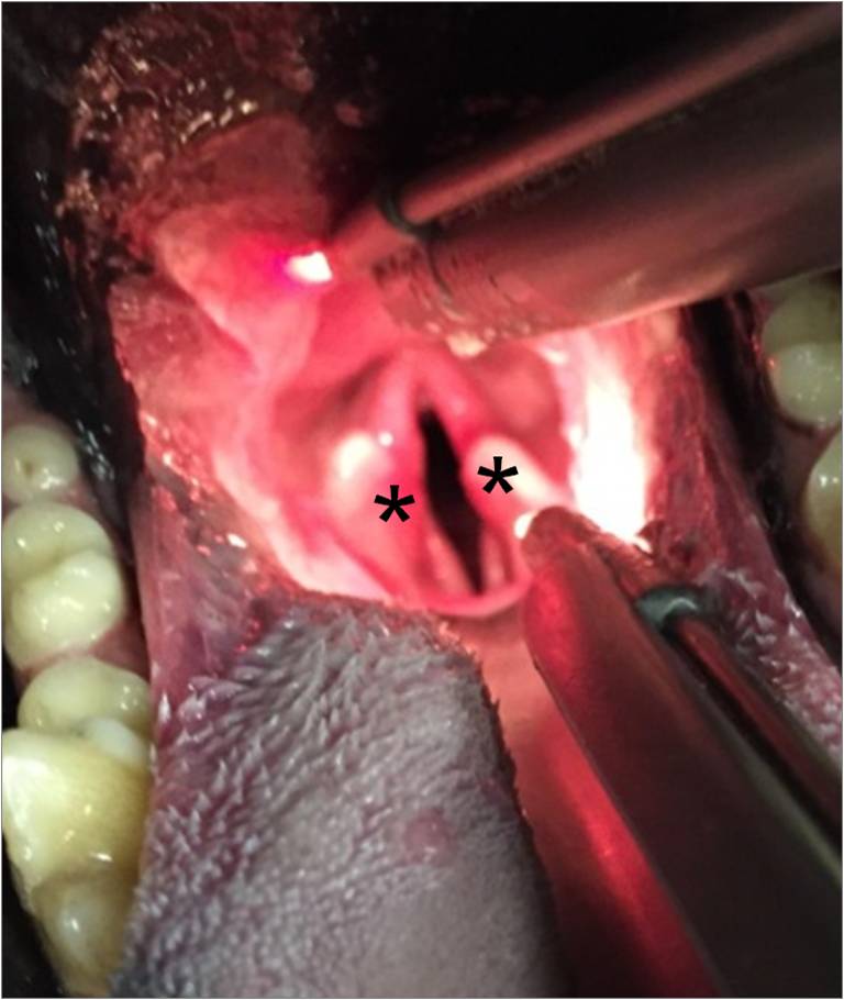
Figure 1. Intra-oral examination of the larynx in a dog with laryngeal paralysis under light sedation. The laryngeal opening into the trachea (black space) is narrowed and the arytenoid cartilages (asterisks) failed to open during inhalation.
Figure 2. A video showing a laryngeal exam of a dog performed under light sedation. Examination showed that both of the arytenoid cartilages of the larynx were paralyzed and did not open during breathing, creating an obstruction of the airway. This confirmed the diagnosis of laryngeal paralysis.
(Video courtesy of Dr. Steven Fernández)
Treatment
Conservative treatment of this condition involves weight management, exercise restriction, avoiding high ambient temperatures, sedatives, and stress reduction. Unfortunately, this condition is progressive and can cause death by suffocation and hyperthermia if not treated when the dog begins to show clinical signs. Any animal with difficulty breathing should be taken to a veterinarian or animal emergency hospital immediately.
Surgical treatment is recommended in dogs diagnosed with laryngeal paralysis. Older surgical procedures involved removing a portion of the larynx in order to enlarge the laryngeal opening; these procedures resulted in an unacceptably high complication rate. The most common surgery currently performed is the unilateral arytenoid lateralization (UAL) – also known as a “tie-back”. The UAL procedure involves tying heavy gauge sutures between the arytenoid and cricoid cartilages on one side of the larynx. This results in one side of the larynx being “tied open” so that airflow is less obstructed and the dog can breathe easier (Figure 3).
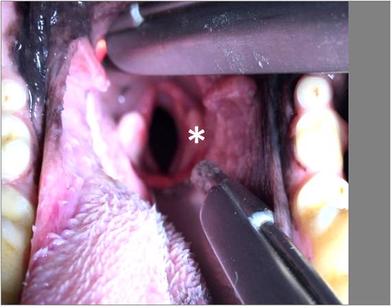
Figure 3. Intra-oral examination of the larynx in the dog in Figure 2 after unilateral arytenoid lateralization showing an enlarged laryngeal opening (black space). One arytenoid cartilage (white asterisk) has been “tied back,” or lateralized.
Potential Post-operative Complications
Clinical signs of difficulty breathing are improved in approximately 90% of dogs after UAL. There are numerous short and long-term possible complications associated with the surgery. Suture breakage, infection of the incision, persistent coughing, or respiratory signs are the most common short-term complications that can be seen.
The most serious complication associated with UAL is aspiration pneumonia, which is an infection in the lungs resulting from food material aspirated during eating or regurgitation. Aspiration pneumonia can be life-threatening and requires immediate medical treatment. Since a portion of the larynx is permanently tied open, dogs after UAL are permanently predisposed to aspiration pneumonia (8-21% lifetime risk). Symptoms of aspiration pneumonia are coughing, difficulty breathing, episodes of respiratory distress (which can be seen as pale/blue gums, gasping for air or using the muscles of the abdomen to breathe), lethargy, inappetance, exercise intolerance, and/or a fever. If any of these signs develop, the patient should see a veterinarian right away. The UAL procedure is not recommended in dogs that have megaesophagus because the risk of aspiration pneumonia is very high. Food material that is regurgitated through the mega-esophagus can easily be aspirated into the lungs since the laryngeal opening is permanently tied open on one side. In that case, a permanent tracheostomy (a permanent opening between the trachea and the skin) may be considered.
Post-operative Care:
Attentive post-operative care for dogs that had the UAL procedure is crucial for a good outcome since one portion of their airway has been tied open permanently. Immediately after surgery it is important to restrict their exercise for 3-4 weeks to allow the incision and surgical site to heal adequately. Soft food formed into small meatballs should be given for the first week in an elevated dish 3-4 times daily during the initial healing period. Then the normal diet can be gradually re-introduced and the frequency of the meals can decrease back to the dog’s normal routine. Anti-inflammatories and pain medication are important to decrease any swelling that may occur in the larynx and provide comfort. Sedatives are prescribed for several weeks as dogs become acclimated to their ‘new’ airway; they may be necessary long-term in dogs that are overly anxious or being too active.
Aggressive physical activity should be avoided permanently as to not put excessive strain on the airway; light play and a moderate amount of physical activity is allowed as long as it is well tolerated and no episodes of respiratory distress are seen (described above). Swimming is allowed after 3-4 weeks (once the surgical site is healed) as long as dogs are monitored closely and keep their heads above the water (to prevent aspirating pool water into the open airway). If there is any concern that a dog is swallowing or inhaling pool water they should not be allowed to swim.
A harness around the body, instead of a neck leash, should be used for the rest of life, so that pressure isn’t applied to the neck area. During warm and hot months, outside activity should be minimal. There must always be continuous free access to water and an easily accessible cool place to rest – ideally indoors in air conditioning.
Prognosis
Since laryngeal paralysis is usually a symptom of a systemic neurologic disease that affects the whole body, additional symptoms of weakness, inability to walk, and difficulty swallowing often develop over time. The laryngeal UAL (tie-back) surgery can provide relief from difficulty breathing until other symptoms progress in severity. Most dogs with the UAL surgery have a good quality of life for 1-2 years after surgery.
Timothy A. Geis, DVM
Comments Closed